Cross Section Of A Bone Diagram - Cross-section of the Long Bone. This simply involves placing a section of the bone on the microscope stage and viewing the. As the names suggest compact bone looks compact and the spongy bone looks like skull bone is a flat bone. Compact bone cross section courtesy: Only the outer shell of electrons of each atom is drawn. Photos of hyomandibular surface showing blocks of tesserae (brownish).
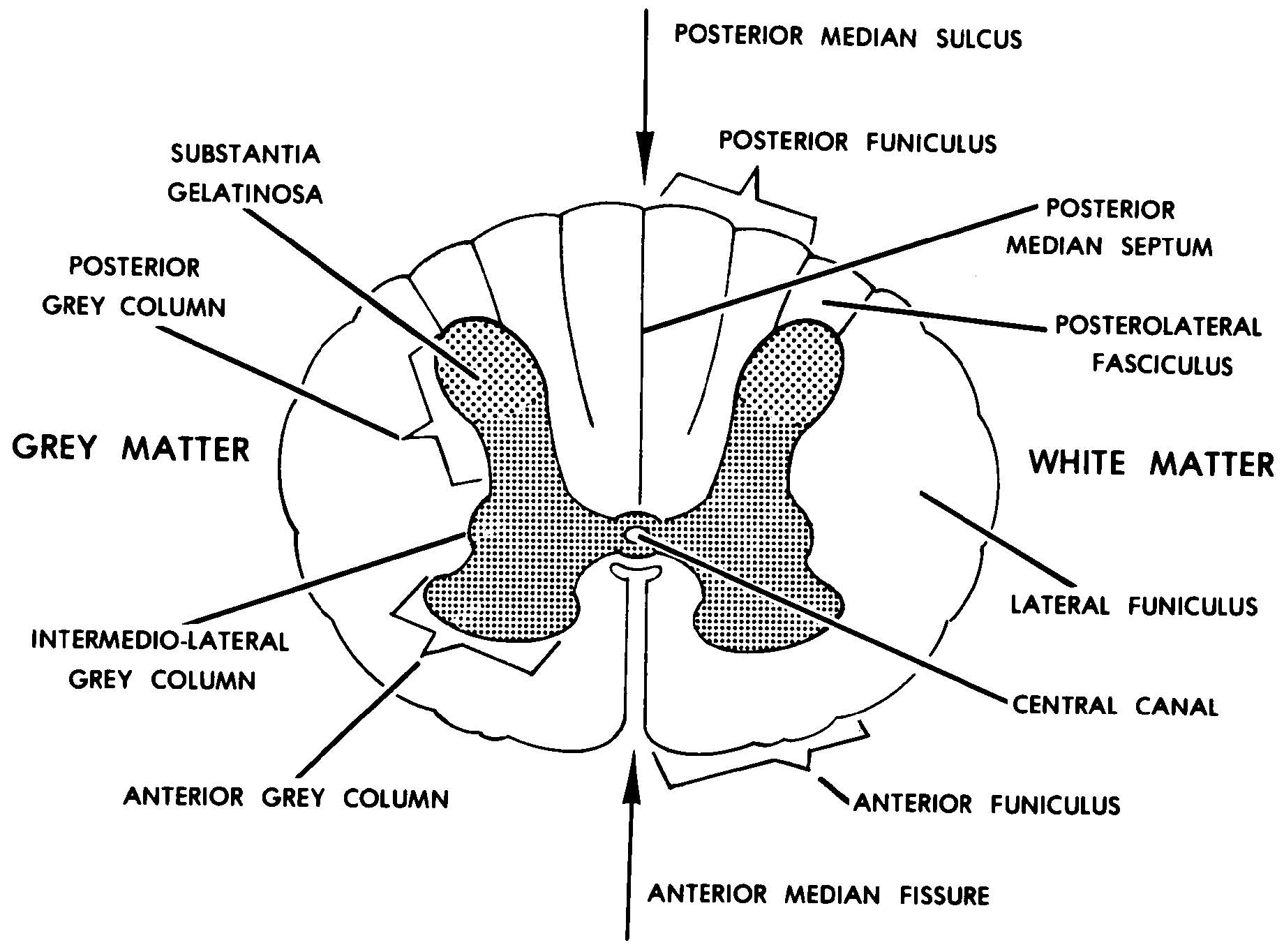
A long bone has two main regions: Cord spinal cross section spine cervical diagram education science anatomical anatomy atlas back body bone care column disc disease foramen fracture grey health healthcare healthy human illustration infographic injury matter medical nerve nervous pain part physiology poster process skeletal skeleton. Shows the ionic bonds between ions by the loss and gain of electrons. They support the body structurally, protect our vital organs, and allow us to move. Two types of bone tissues in cross section of a long bone :
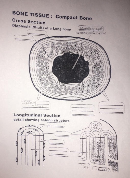
Images 11. Nervous System | Basic Human Anatomy from brooksidepress.org Medically reviewed by the healthline medical network — written by the healthline editorial team — updated on january 20, 2018. Only the outer shell of electrons of each atom is drawn. Diagram with articular cartilage, marrow, medullary cavity and periosteum. We can see there are two layers of compact bone here. Vector illustration scheme of bone cross section. Two types of bone tissues in cross section of a long bone : Cord spinal cross section spine cervical diagram education science anatomical anatomy atlas back body bone care column disc disease foramen fracture grey health healthcare healthy human illustration infographic injury matter medical nerve nervous pain part physiology poster process skeletal skeleton. A long bone has two main regions:
Whereas a long bone has only one layer of compact bone (see fig 1).
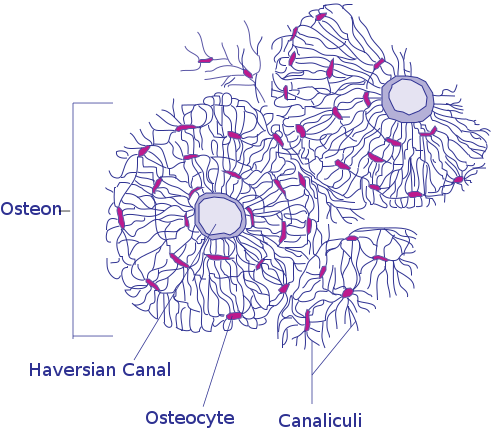
Bones in the foot diagram. They build the entire picture, improve your understanding, consolidate the information and facilitate recall. Bone is found in the shafts of long bone and consists of various cylindrical units named as haversian system 47. Only the outer shell of electrons of each atom is drawn. As shown in figure 2. Spongy bone is composed of trabeculae that contain the osteocytes. Blue square delimits one tesseral block (after dean and summers, 2006). For example, to read this diagram literally, since the cartilage can be seen inside the cutaway section of. Explaned distal and proximal epiphysis. Hope you enjoy and please. Bone marrow is the soft, highly vascular and flexible connective tissue within bone cavities which serve as the primary site of new blood cell production or bone marrow is the primary source of pluripotent stem cells that give rise to all hemopoietic cells (blood cells) including lymphocytes. It seems confusing and misleading. Two types of bone tissues in cross section of a long bone :
The leg bone's connected to hip bone. This is a short tutorial using blender 2.8 that shows how to create a bone cross section and using images to create the textures. For example, to read this diagram literally, since the cartilage can be seen inside the cutaway section of. As the names suggest compact bone looks compact and the spongy bone looks like skull bone is a flat bone. A long bone has two main regions:
Solved: BONE TISSUE: Compact Bone Cross Section Diaphysis ... | Chegg.com from media.cheggcdn.com Bone anatomy sketches 12 photos of the bone anatomy sketches , bone. Only the outer shell of electrons of each atom is drawn. Electrons in one atom will be represented as crosses, whilst electrons in the other atom will be represented as circles, indicating which ion gains or. (b) in this micrograph of the osteon, you can clearly see the concentric lamellae and central canals. The periosteum contains many strong collagen fibers that are used to firmly anchor. The large dark spots are passages for blood vessels and nerves. Compact bone cross section courtesy: Vector illustration scheme of bone cross section.
The periosteum contains many strong collagen fibers that are used to firmly anchor.
The large dark spots are passages for blood vessels and nerves. Cross section of bone diagram. Each system contains the main advantage of this method is the enhancement in electrospinnability of a less spinnable material with the help of a highly spinnable. (micrograph provided by the regents of university of michigan. Only the outer shell of electrons of each atom is drawn. Learn all about the major systems of the body from the integumentary system to the nervous system using our fun and engaging human body unit bundle, complete with a guiding powerpoint. Jump to navigation jump to search. Blue square delimits one tesseral block (after dean and summers, 2006). As shown in figure 2. Bone anatomy sketches 12 photos of the bone anatomy sketches , bone. Two types of bone tissues in cross section of a long bone : The periosteum contains many strong collagen fibers that are used to firmly anchor. It seems confusing and misleading.
Crystals of apatite and a typical section of a long bone of a mammal cut approximately parallel to the fibril axes is shown in figure 2. Shows the ionic bonds between ions by the loss and gain of electrons. Looking at a bone in cross section, there are several distinct layered regions that make up a bone. As shown in figure 2. Bone is a biologically generated composite material comprised of two major structural components:
File:Transverse Section Of Bone.svg - Wikimedia Commons from upload.wikimedia.org Shows the ionic bonds between ions by the loss and gain of electrons. This simply involves placing a section of the bone on the microscope stage and viewing the. Cord spinal cross section spine cervical diagram education science anatomical anatomy atlas back body bone care column disc disease foramen fracture grey health healthcare healthy human illustration infographic injury matter medical nerve nervous pain part physiology poster process skeletal skeleton. Thus, the motions of the body and its parts, all the way from the lunge of the football player to the delicate manipulations of a handicraft artist or of the use of complicated instruments by a scientist, are made. The large dark spots are passages for blood vessels and nerves. Diagram with articular cartilage, marrow, medullary cavity and periosteum. Medically reviewed by the healthline medical network — written by the healthline editorial team — updated on january 20, 2018. Jump to navigation jump to search.
Cross section through an model of an fracture in the neck of the femur (thigh bone).
Also, they provide an environment bones are mostly made of the protein collagen , which forms a soft framework. Compact bone cross section courtesy: Shows the ionic bonds between ions by the loss and gain of electrons. Bone is found in the shafts of long bone and consists of various cylindrical units named as haversian system 47. We can see there are two layers of compact bone here. Uc, uncalcified to calcified cartilage interface; Thus, the motions of the body and its parts, all the way from the lunge of the football player to the delicate manipulations of a handicraft artist or of the use of complicated instruments by a scientist, are made. Two types of bone tissues in cross section of a long bone : (b) in this micrograph of the osteon, you can clearly see the concentric lamellae and central canals. Learn all about the major systems of the body from the integumentary system to the nervous system using our fun and engaging human body unit bundle, complete with a guiding powerpoint. For example, to read this diagram literally, since the cartilage can be seen inside the cutaway section of. They support the body structurally, protect our vital organs, and allow us to move. Blue square delimits one tesseral block (after dean and summers, 2006).
Diagram with articular cartilage, marrow, medullary cavity and periosteum cross section of a bone. These solutions assume a circular cross section whereas long bones generally are far less uniform.
Share :
Post a Comment
for "Cross Section Of A Bone Diagram - Cross-section of the Long Bone"

Post a Comment for "Cross Section Of A Bone Diagram - Cross-section of the Long Bone"